This comprises the nose, the pharynx, the Wind-pipe, the Lungs, and the Diaphragm.
The nose is a cavity with an anterior and a posterior opening. There are two cavities and two front and back openings, one for each side of the body—separated medially by means of a septum. The external aspect presents, among others one ala on each side, an ala being the expanded mobile lower part of internal surface of nose. The Septum or middle partition is in part bony and in part cartilaginous. From the lateral wall of the nasal cavity, are suspended the superior, the middle and the inferior turbinated bones. The whole inside of nose (the nasal cavity or fossa) is lined by mucous membrane and the outer wall of each cavity communicates with the hollow in the upper cheek-bone (superior maxilla) called the Antrum of Highmore. Where the nasal cavities open into the upper part of what forms the back of the mouth (pharynx), it is called the posterior nares.
Pharynx is that cavity which forms behind the nose and mouth and freely communicates with them. This is the junction-station as it were of five important structures, viz;
Anteriorly and in its upper part—of the two nasal cavities;
Laterally—of the two Eustachian Tubes;
Centrally and in front of the mouth;
Inferiorly—of the wind-pipe in front; and the œsophagus behind.
For the purpose of anatomical convenience, the upper part of pharynx is called naso-pharynx, the central part oral pharynx, and the lower part laryngeal pharynx. In the oral part are situated the tonsils, one on each side.
Larynx is the organ of voice, arising where the pharynx ends viz., from below the back of tongue. It has several cartilages. (1) The Thyroid Cartilage, quadrilateral in shape, one on each side, and situated prominently in the front part of the neck, where it projects as pomum adami. Inside them lie the vocal cords. (2) Cricoid cartilage shaped like a signet-ring, is situated below the thyroid cartilage. (3) On the superior border of the cricoid cartilage, lies the Arytenoid cartilage, one for each side, and to each of them is attached a vocal cord. (4) Epiglottis is situated at the root of tongue, anterior to the upper aperture of larynx. It is shaped like a leaf. The larynx is lined throughout by mucous membrane, continuous with that of pharynx above, and trachea below.
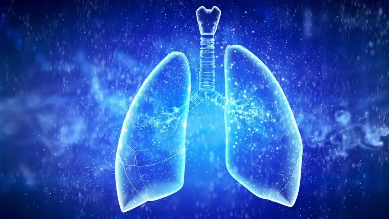
Trachea and Bronchi—Trachea begins at lower border of cricoid cartilage. It ends by dividing into two bronchi, opposite enteral angle. Each bronchus enters the lung, relating to its side, where it divides into smaller and smaller branches (bronchiole) and is lined throughout by mucous membrane. The final termination of a bronchiole is into air-sacs which are called alveoli.
The Thorax or cavity of chest is subdivided into two pleural cavities and an intervening region, the mediastinum. The latter is occupied principally by the heart. Each lung is covered by a very delicate serous sheet, which is called the pleura.
Lungs are soft, spongy and elastic; they are full of air inside sacs. They are pyramidal in shape with the tapering apex projecting into the sides of the neck, the hollowed-out base resting on the diaphragm. The left lung has two lobes and the right has three lobes, the lobes being separated by fissures. The two lungs fill up practically four-fifth of the chest and are covered by the pleura, a serous covering. The ultimate unit or component of the lung is a bunch of air-vesicles or cells, attached to minute bronchiole; such a piece looks like a bunch of grapes around a single stalk and it is into these air-vesicles that ultimately air enters. Each such piece (a sheath of air-vessels attached to a bronchiole) is called an infundibulum, and is surrounded by a network of capillaries.
Diaphragm is the musculo-tendinous partition between the thorax and abdomen. It is attached, in front, to the back part of the xiphoid appendix and to the inner surface of the lower six costal cartilages; behind, to the bodies of the upper three lumbar vertebrae; and on the lateral aspects, to the costal arches. It supports on itself the lungs and the heart; and in the abdomen, immediately below it, lie on the left side, the spleen and stomach and on the right, the liver. Its two pillars give support to two kidneys.
Through it pass, from thorax into abdomen, the œsophagus and aorta; from abdomen into the thorax, pass the inferior vena cava and the thoracic duct and other nerves and vessels.
Comments are closed.